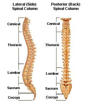
The spine is made up of lots of small bones called vertebrae or vertebral bodies.
The spine consists of
- 7 cervical vertebrae within the neck
- 12 thoracic vertebrae – which are attached to the ribs on each side to form the rib cage
- 5 lumbar vertebrae in the lower back
- The sacrum – which is joined to the pelvis via the sacro-iliac joints.
- The coccyx – which is below the sacrum and is sometimes called the ‘tail-bone’.
Looking at the spine from the back it appears to be straight.
From the side there are three curves; in the cervical spine the neck curves backwards (lordosis), in the thoracic spine it curves forwards (kyphosis). Again in the lumbar spine it curves backwards (lordosis).

The diagram shows the different parts of the vertebral bones.
At each level of the spine, except for the two cervical vertebrae at the top and the sacrum and coccyx, the basic anatomy of the vertebral bones are the same – although the size and shape of the different vertebrae varies between the different areas of the spine.
Each vertebra consists of the vertebral body at the front, the pedicles at the sides and the lamina at the back of the spine. This creates a bony canal (a space) in the centre for the spinal cord. At the back of the vertebrae there are a pair of facet joints which are the joints between the adjacent vertebra above and below.
At each level of the spinal column there a pair nerve roots that exit through a hole in the vertebra (the ‘foramen’). Within the cervical and lumbar spine these nerve roots join together to form the cervical and lumbar plexuses, which supply the power and sensation the arms and legs respectively.
Each individual nerve root supplies a specific area of the skin (dermatome) and specific muscles (myotome).
Between each vertebra there are three joints – the disc between the bodies at the front, and two smaller facet joints at the back of the spine. This constitutes a “motion segment” of the spine. The amount of movement within a given motion segment varies depending on which area of the spine, with greater movement possible in the cervical and lumbar regions.
The vertebral bodies are separated by cushions called intervertebral discs. A disc is made of special cartilage and acts like a shock absorber between the bodies. The disc is made of two parts – the outer layers form the annulus fibrosis, fibers arranged in a circular pattern. In the centre of the disc is the nucleus pulposis, which has a more gelatinous form. When the annulus tears central nucleus may prolapse through the annulus and press on the nerve root.
The upper two cervical vertebrae have a different structure compared with the other vertebral bodies.
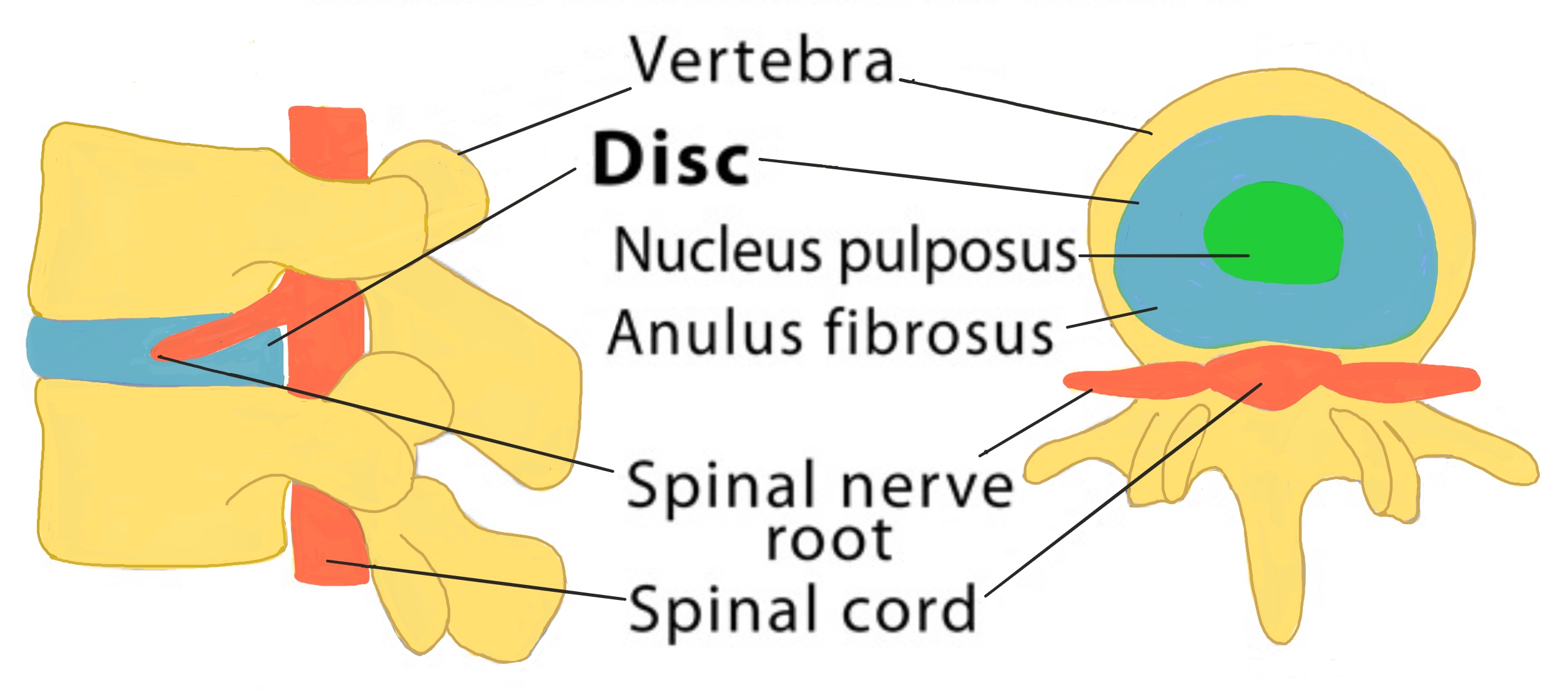
Diagram shows the side view of the vertebrae and cross-section through the intervertebral disc.