An MRI or Magnetic Resonance Imaging Scan can give a very clear picture of the structure of the spine, there by allowing your surgeon to look at the nerves, bones and soft tissues of your spine. The scan is extremely sensitive, but is only useful when the findings are interpreted in conjunction with a full history of the spinal condition and a thorough examination.
CT images show bones, their shape, position and anatomy. CT (sometimes called CAT) scanners use radiation (X-rays) to form an image and can be used in conjunction with MRI scans for both making a diagnosis and for planning an operation.
What is an MRI scan?
The MRI Scan uses magnetic and radio waves, meaning that there is no exposure to X-rays or any other damaging forms of radiation. Radio waves stronger than the magnetic field of the earth are then sent through the body. This affects the body’s atoms, forcing the electrons into a different position. As they move back into place they send out radio waves of their own. The scanner picks up these signals and a computer turns them into a picture.
Are MRI scans safe?
There are no known dangers or side effects connected to an MRI scan, however there is a small theoretical risk to the foetus during the first 12 weeks of pregnancy and ideally an MRI scans should be avoided during this period.
What is an MRI scan with STIR sequence?
MRI scans with a STIR sequence are performed to help identify infection or cancers in the spine – theses are seen as areas of high signal (bright white). From a patients perspective, there is no difference in how the scan is performed.
Do I need a scan?
The vast majority of patients with back pain can be treated following a careful history and examination of the spine. An MRI or CT scan is not usually required, as this will not change the treatment for their back pain. A scan is however useful when there are symptoms of persistent nerve root irritation, causing pain in the arm or the leg, or for patients with severe persistent back pain in which injection therapy or surgery is being considered. On rare occasions back pain is caused by a serious condition such as a tumour, fracture or infection of the spine. Usually these conditions can be excluded with a thorough history and examination, however if a patient has “Red Flag Signs” then an MRI or CT scan is required to exclude a serious condition.
The scan does not tell us where back pain comes from, but may help localise the source of the pain and allow treatment to be directed to the appropriate area of the spine.
However remember “We treat patients not a scan.”
What to expect if you are sent for an MRI scan
Before having an MRI scan you will be asked to complete a safety questionnaire. Some patients may not be suitable for a scan, reasons for this include people who are fitted with a cardiac pacemaker or have metal fragments in their eyes. These patients may then require a CT scan of the spine instead.
Patients lie inside a large cylinder-shaped magnet, usually for about twenty minutes, while the scans are being made. The scanner makes a loud banging noise, which might be unpleasant, but usually music or ear defenders are available to limit this affect.
Some people may feel claustrophobic during the test. Patients who are afraid this might happen should talk to the doctor beforehand, who may give them some medication to help them relax, or arrange for the MRI scan to be performed in a larger open scanner, although the images may be of a slightly poorer quality. Often, in the modern scanners, people are able to enter feet first and the scanners are much shorter, both factors greatly reduce the feelings of claustrophobia. If you can keep very still in the scanner, the picture quality is better.


CT Scans
CT (Computerised Tomography) scanners look like a big polo mint and are a lot quicker than MRI scans. CT scans are produced by using radiation (X-rays) to form an image. They are used to look at bones, their shape, position and anatomy and can be used in conjunction with MRI scans when planning an operation. In the spine, CT scans are also good at looking post-operatively at the position of screws and metalwork and to assess fusions. CT scans are also used for patients who cannot have MRI scans, for example those patients who have non-MRI compatible pace-makers, metal clips in the brain or metal fragments in the eyes.
CT SPECT Scans
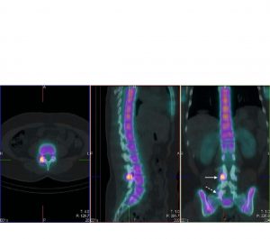
A CT SPECT scan is a 3-D image made by combining 2 separate images; a CT scan and a SPECT scan. The fused images can sometimes provide more accurate information about the anatomy and function of the area being scanned in difficult or unusual problems. For example, in areas such as the spine, it can be difficult to determine from one form of imaging alone whether the issue lies in a fusion site or the adjacent facet joints. The combination of SPECT and CT images may provide further information to add confidence in identifying and localizing the problem.
Single Photon Emission Computed Tomography (SPECT) images are produced following an injection of radio-labelled dye (which contains a small amount of radioactivity). The images produced have coloured areas “hot spots”, indicating how an area of the body is functioning and may identify potential sources of pain.
The CT and SPECT images are obtained while you lie on the same imaging bed at the Same appointment . During the SPECT procedure a special camera rotates in a 360 degree arc allowing for reconstruction of 3-D images. For the CT scan the bed then slides through the centre of the scanner. Again, the scanner rotates over a 360 degree arc allowing for 3-Dimage reconstruction. Currently there are not many CT SPECT scanners in the UK.
However, it is important to remember these are NOT “pain maps” and as with all imaging, results need to be used in combination with the history and examination of a patient.

The CT SPECT images show increased tracer uptake at the right L3/4 facet joint, and to a lesser extent the right L5/S1 facet joint. If the patients symptoms correspond with the results of the scan, it is likely that the source of the pain is the right L3/4 facet joint.


